
Things You Need to Know About Histology Lab Process
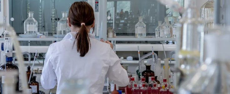
This report illustrates the six stages that the tissue biopsy goes through in the pathology laboratory. The pathologist seems to be waiting on a slide in a histology laboratory. Hence, you can read more reference on https://www.journals.elsevier.com/clinica-chimica-acta/news/euromedlab-2019-abstracts to know more about medical lab facts.
The Histology Lab Process

Probably the last thing you’re worried about while you’re waiting for the doctor to tell you something is, I wonder what’s in my biopsy scheduled right now. No, you want the lab results from the health care system, and you want them now because life can change dramatically with just one call from your doctor. The histology lab is where all biopsies are prepared for analysis by a pathologist. I want to describe this process that many surgical studies will go through.
A biopsy is a tissue taken from a living body to be analyzed to determine the presence, activation, or extension of the disease. The same thing happens microscopically with the small piece of tissue called a biopsy. The strings of protein molecules begin to appear almost immediately after removing the tissue. We call it tissue autolysis. Think back to the cut umbilical cord. One thing you could do to prevent it from dissolving would be to keep a match lit at the frayed ends to dissolve the fibers. To stopped tissue autolysis, the inserted biopsy into a tissue processor that uses 10% neutral buffered formalin to pass through the ends of the amino acids and prevent further degradation.
The Result of Histology Lab Process
When the surgical sample arrives at the histopathology laboratory, it transfers to a raw station. There it is thoroughly examined for diseases, measured, and explained in another way. Each day, a histologist selects each of the extrapolated cassettes and inserts them into a tissue processor. Depending on these individual pieces’ size, fabric processing can take from four weeks to thirteen hours. The fabric pieces move the veins through another set of chemical reagents. Formalin fixes or stops the fabric from breaking. Three parts or alcohol markers slowly dry the material. Xylene removes the alcohol from the tissue.
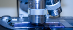
The tissue is removed from the tape and placed in a mold filled with liquid kerosene, the tape on which the person’s hospital number prints inserted into the mold. The hot kerosene flows from the embedding station into the mold. Microtomy, thin sections of biopsy tissue are reduced and placed on glass slides. The kerosene block transfers into an instrument called a microtome. The block removes from the carriage, which moved to a blade. Each time the cube moves the edge, it advances four or even four micrometers. This is the thickness of a block of fabric. Considering that the fabric continues to carry on the blade, several segments produce a very long strip.…